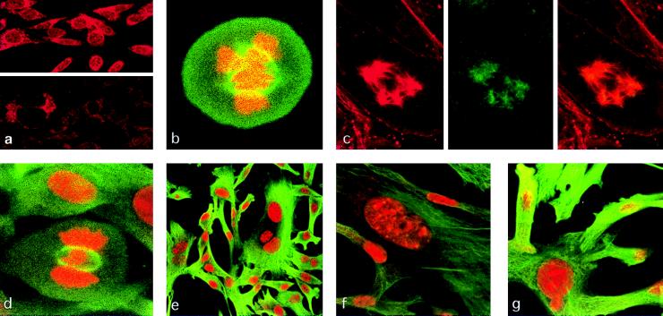
Figure 4.
Mitotic spindles, microtubule organizing centers, and nuclear morphology of SL6 cells. (a) NIH 3T3 (Upper) and SL6 (Lower) cells stained by anti-TSG101 antibody. (b) Cell stained by anti-tubulin antibody (green) and propidium iodide to detect DNA (red). (c) Tubulin (Left), TSG101 (Center), or both (Right) staining in an SL6 cell containing five centrosomes; other metaphase cells in this preparation showed similar abnormalities. (d) “H”-shaped metaphase DNA (red) and tubulin-stained mitotic spindle (green) in SL6 cell. (e–g) Nuclear abnormalities in SL6 cells, stained for tubulin (green) and DNA (red). All photographs were made by using confocal microscopy.

