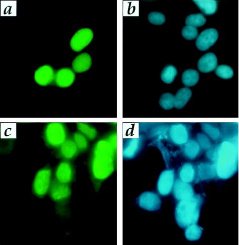
Figure 1.
Immunofluorescence of menin-transfected HEK-293T cells with menin and myc epitope antibodies. Twenty-four hours after transfection with pcDNA3.1-menin, cells were processed for immunofluorescence with menin antibody (KC27) or with myc antibody followed by fluorescein isothiocyanate-conjugated secondary antibody detection. Immunofluorescence pattern with menin antibody (a) and the DAPI staining (b) showing the nuclei from the same cells. Immunofluorescence with myc antibody (c) and the DAPI staining (d) showing the nuclei from the same cells. Note that not all cells are positive, because this is a transient transfection. Endogenous levels of menin are not detectable above background.

