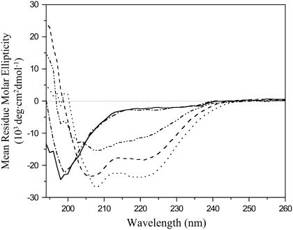
FIGURE 7.
CD spectra of HIV-1 gp41 fusion domain in solution, bound to DPC micelles, and bound to lipid bilayers composed of POPC/POPG (4:1). (Solid line) 0.1 mM P23H8 in 5 mM HEPES, 10 mM MES buffer, pH 7.4. (Dash-dotted line) Same with 0.2 M NaCl added. (Dash-dot-dot line) Same with 1 M NaCl added. (Dashed line) 0.1 mM P23H8 bound to 10 mM DPC micelles. (Dotted line) 0.1 mM P23H8 bound to 10 mM POPC:POPG (4:1) bilayers. All spectra were recorded at 25°C.

