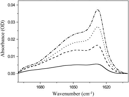
FIGURE 8.
ATR-FTIR spectra of HIV-1 gp41 fusion domain bound to planar-supported lipid bilayers composed of POPC/POPG (4:1) at different protein concentrations. (Solid line) Ten micrograms per milliliter P23H8 added to bilayer. (Dashed line) Twenty micrograms per milliliter P23H8 added to bilayer. (Dotted line) Thirty micrograms per milliliter P23H8 added to bilayer. (Dash-dotted line) Forty micrograms per milliliter P23H8 added to bilayer. All spectra were recorded at room temperature. The spectrum of the pure bilayer recorded before the first protein addition was subtracted from each spectrum.

