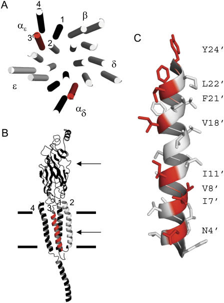
FIGURE 1.
M3 of the α-subunit. (A) Torpedo AChR transmembrane domain, viewed from the synapse (PDB code 2bg9 (7)). M3 in the two α-subunits is red. In all subunits M2 lines the channel and M4 is at the periphery. (B) αɛ-subunit, viewed from the membrane. The upper and lower arrows mark the transmitter binding site and the presumptive gate at the M2 equator; the thick horizontal lines mark approximately the membrane. (C) The M3 helix of the αɛ-subunit, viewed from the membrane. The residues that were mutated (red) mostly face the membrane. In mouse, the αM3 sequence (24′–1′) is YMLFTMVFVIASIIITVIVINTHH (Table 1). In Torpedo αM3 has two differences, at 7′ (I→V) and 14′ (A→S).

