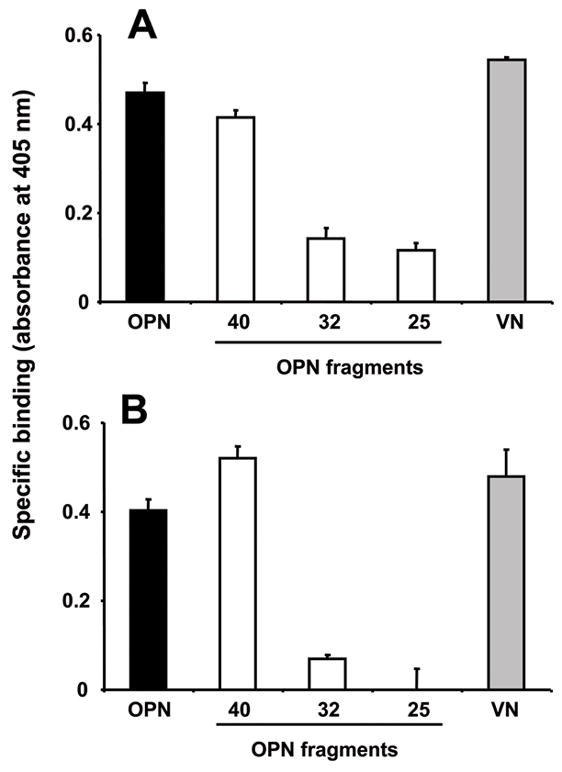
Figure 1. Binding of Eap to osteopontin.

(A) Binding of full-length OPN (filled bar), the OPN fragments, (40-kD, 32-kD and 25-kD) (open bars) or vitronectin (gray bar) to immobilized Eap is shown. (B) Binding of Eap in solution to immobilized full-length OPN (filled bar), the OPN fragments, (40-kD, 32-kD and 25-kD) (open bars), or vitronectin (gray bar) is shown. Specific binding is expressed as absorbance at 405 nm. Data are mean±SD (n=3) of a typical experiment; similar results were observed in three separate experiments.
