
Figure 4. Reduction of MDA-MB-231 metastasis to the bone by Eap.

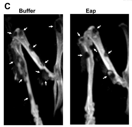
The metastasis of MDA-MB-231 cells to the bone was studied in buffer- or Eap-treated mice. (A) The total volume (in mm3) of metastases was estimated by volumetric CT and (B) the total metastasis count in vertebrae and extremities was counted at days 21 and 35 after the initial intracardiac injection of MDA-MB-231 cells. Data are mean±SD (n=5 mice/group). (C) Representative CT images from the lower extremities of a buffer-treated mouse and an Eap-treated mouse 35 days after initial intracardiac inoculation of MDA-MB-231 cells. Metastatic osteolytic lesions are depicted by arrows. *: P<0.05, as compared to buffer; **: P<0.01, as compared to buffer.