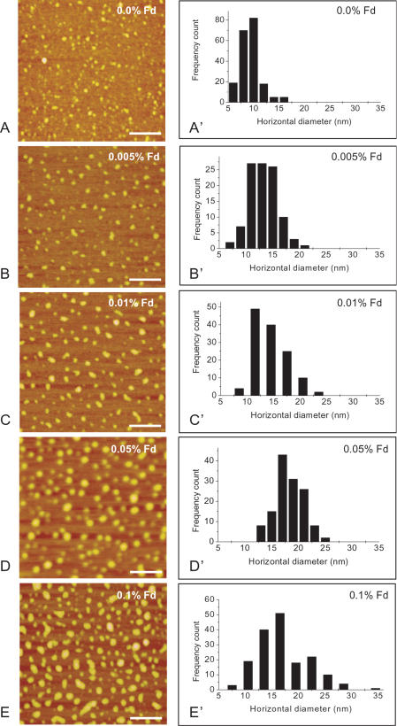
Figure 1. AFM Images of neuronal tau in the presence of formaldehyde at different concentrations.
Neuronal tau (20 µM) was incubated in 50 mM phosphate buffer (pH 7.2) containing formaldehyde at different concentrations as indicated at 37°C over night (A–E). Aliquots were taken and diluted to the desired concentration using the phosphate buffer, and the samples were dropped onto mica surfaces and dried in air before observed under the atomic force microscope. The frequency counts of the horizontal diameters (explained in the text) of the protein particles in images A–E were shown in A'–E', respectively. The scale bars equal 100 nm.

