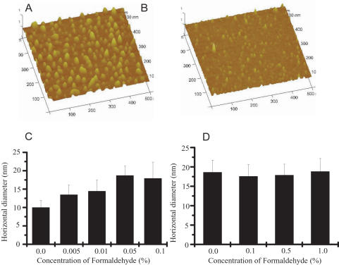
Figure 2. Three-dimensional images and horizontal diameters of tau at different concentrations of formaldehyde.
The data using for regenerating three-dimensional images of neuronal tau in the presence (A) and absence (B) of formaldehyde solution (0.1%) are the same used for Figures 1A and E. Change in the horizontal diameter of the tau protein particles on mica surface at different concentrations of formaldehyde is depicted in C. Change in the horizontal diameter of BSA was used as control (D).

