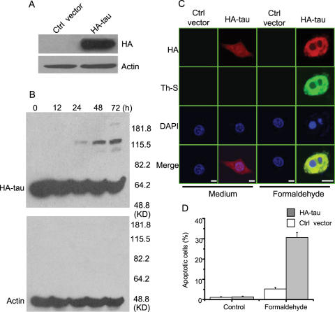
Figure 10. Toxicity of tau aggregation to HEK 293 cells in the presence of formaldehyde.
HEK 293 cells were cultured and transient with HA-tau or the control vector, followed by incubation with 0.0002% formaldehyde for 72 h. Immunolabelling with the monoclonal antibody HA was used to visualize tau expression (A) and tau aggregation in the presence of 0.0002% formaldehyde for different times (B). β-actin was used as a protein loading control. Immunostaining detected tau aggregation in 293 cells after treated with formaldehyde for 72 h (C, Bar: 25 µm). Other conditions were similar to SY5Y cells shown in Figure 9. Quantitative analysis is presented in D. Apoptotic cells are characterized by nuclear condensation and diffraction. At least 200 apoptotic cells were counted in each experiment.

