
Fig. 4.
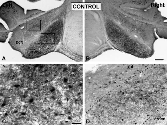
Low- (A,B) and high- (C,D) magnification digital images illustrating calretinin immunostaining in the PVCN and DCN in control ferrets. In the PVCN, a dense calretinin immunostained neuropil and immunostained cells were evident throughout the nucleus (C). In the DCN, calretinin immunostained neurons were more abundant in the granular cell layer than in the molecular and central region of the DCN (D). AVCN, anterior ventral cochlear nucleus; crdcn, central region of the DCN; DCN, dorsal cochlear nucleus; grcl, granular cell layer; ml, molecular layer; PVCN, posterior ventral cochlear nucleus. Scale bars = 100 μm in B (applies to A,B); 50 μmin C (applies to C,D).