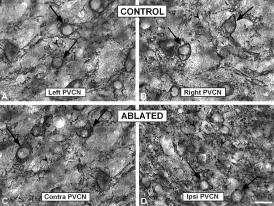
Fig. 9.

High-magnification digital images showing synaptophysin immunostaining in the PVCN in control (A,B) and ablated (C,D) animals. Similar to the anterior ventral cochlear nucleus, large profiles surrounding cell bodies (arrows) were abundant in the PVCN in control and contralateral side in ablated ferrets. In addition, there was a qualitative increase in the number of punctate profiles (arrowheads) in the ipsilateral PVCN (D) compared to the contralateral side (C) and unoperated animals (A,B). PVCN, posterior ventral cochlear nucleus; contra, contralateral; ipsi, ipsilateral. Scale bar = 25 μm in (applies to A–D).
