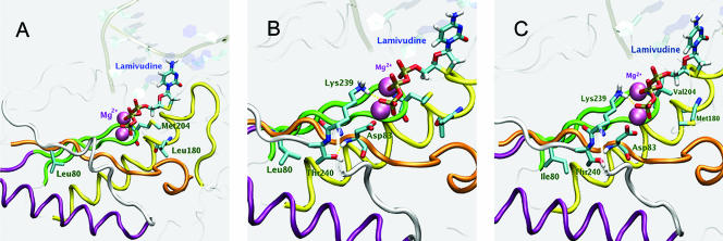
FIG. 4.
(A) Model of the wild-type HBV rt showing the location of residue L80 in relation to residues L180 and M204, which are located in the dNTP-binding pocket. Conserved sequence motifs are colored: A, orange; B, yellow; C, green; D, white. The conserved alpha helix and the chelated magnesium ions are purple. LMV-TP is shown occupying the dNTP-binding pocket. (B) Model of the wild-type rt active site showing the spatial relationships between residues L80, T240, D83, and K239. (C) Model of the dNTP-binding pocket of the active site of LMV-resistant rt showing the location of rtL80I in relation to rtM204V and rtL180M. Note the alteration in the spatial relationship between rtL80I and rtT240 compared to that in panel B.

