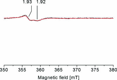
FIG. 2.
Detection of the Fe-S center I from succinate dehydrogenase/fumarate reductase in V. cholerae membranes by EPR spectroscopy. Membranes from the wild-type V. cholerae strain containing the Na+-NQR (black trace) and from the nqrC insertion mutant (red trace) were mixed with 36.4 mM Na2-succinate prior to freezing. Characteristic g values are indicated. For EPR conditions, see the legend to Fig. 1.

