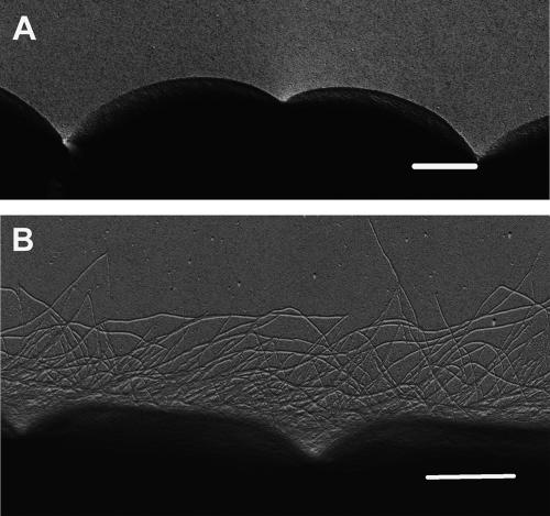
FIG. 2.
Comparison of the cell surfaces of wild-type vegetative filaments and hormogonia. (A) Wild-type vegetative filaments; (B) wild-type hormogonia. Scale bars represent 1 μm. For electron microscopy, platinum wire (2 cm by 0.2 mm) was evaporated onto the surface of each sample by using an Edwards 306A high-vacuum coating unit and samples were viewed on a JEOL1200EX transmission electron microscope at 80 kV.

