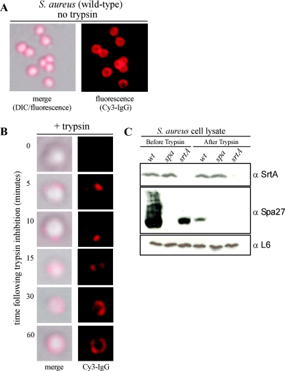
FIG. 1.
Deposition of newly synthesized protein A on the staphylococcal surface. (A) Protein A on the surface of S. aureus RN4220 cells was stained with Cy3-IgG, and DIC or fluorescence images were captured with an Olympus AX-70 fluorescence microscope and Himamatsu CCD camera. (B) Staphylococci were treated with trypsin to remove surface proteins, and the deposition of newly synthesized protein A on the cell surface at indicated times was visualized by staining with Cy3-IgG. DIC and fluorescence images were captured with a CCD camera. (C) Staphylococcal lysates before and after trypsin treatment were precipitated with trichloroacetic acid, washed in acetone, and separated on SDS-PAGE. Samples were subjected to immunoblotting with antibodies against sortase A (α-SrtA), protein A (α-Spa27), or ribosomal protein L6 (α-L6).

