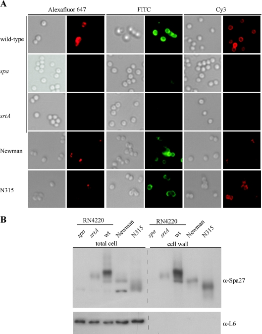
FIG. 2.
Distribution of protein A on the surface of staphylococcal strains. (A) S. aureus RN4220 (wild type) and isogenic spa and srtA mutants were incubated with Alexa Fluor 647-IgG, FITC-IgG, or Cy3-IgG, and DIC or fluorescence images were captured with a Olympus AX-70 fluorescence microscope and Himamatsu CCD camera. As a control for protein A distribution on human clinical isolates, S. aureus Newman and N315 strains were analyzed with the same technology. (B) Total cell extracts or cell wall fractions generated by degradation of murein sacculi with lysostaphin obtained from S. aureus RN4220 (wild-type [wt]) and isogenic spa and srtA mutants or from strains Newman and N315. Samples were separated by SDS-PAGE and then analyzed by immunoblotting with monoclonal antibody specific for protein A (α-Spa27) or with polyclonal antiserum raised against purified L6 ribosomal protein.

