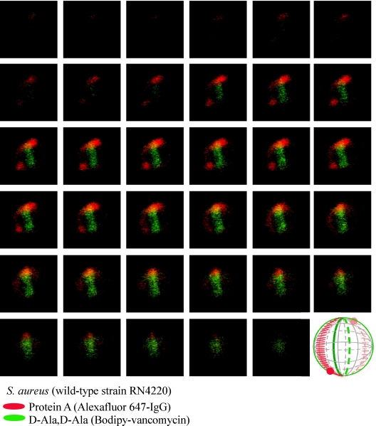
FIG. 3.
Protein A localization in a dividing S. aureus RN4220 cell. Laser scanning confocal microscopy images (z series with 50-nm increments) of S. aureus RN4220. Protein A was labeled with Alexa Fluor 647-IgG (red fluorescence), and cell wall pentapeptide was stained with BODIPY-vancomycin (green fluorescence). Central regions of intense green fluorescence reveal the cross wall, the cell wall layer separating two daughter cells. Each display item is derived from merged images of confocal scans of separate laser line channels. Aggregate data were used to build a three-dimensional model of protein A deposition in the cell wall, which is shown as a diagram in the lower right corner.

