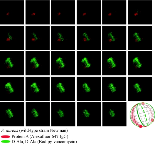
FIG. 4.
Protein A localization in a dividing S. aureus Newman cell. Laser scanning confocal microscopy images (z series with 50-nm increments) of S. aureus Newman. Protein A was labeled with Alexa Fluor 647-IgG (red fluorescence), and cell wall pentapeptide was stained with BODIPY-vancomycin (green fluorescence). Protein A and cell wall images of pentapeptide precursors were collected as described in the legend of Fig. 3. Aggregate data were used to build a three-dimensional model of protein A deposition in the cell wall, which is shown as a diagram in the lower right corner.

