FIG. 7.
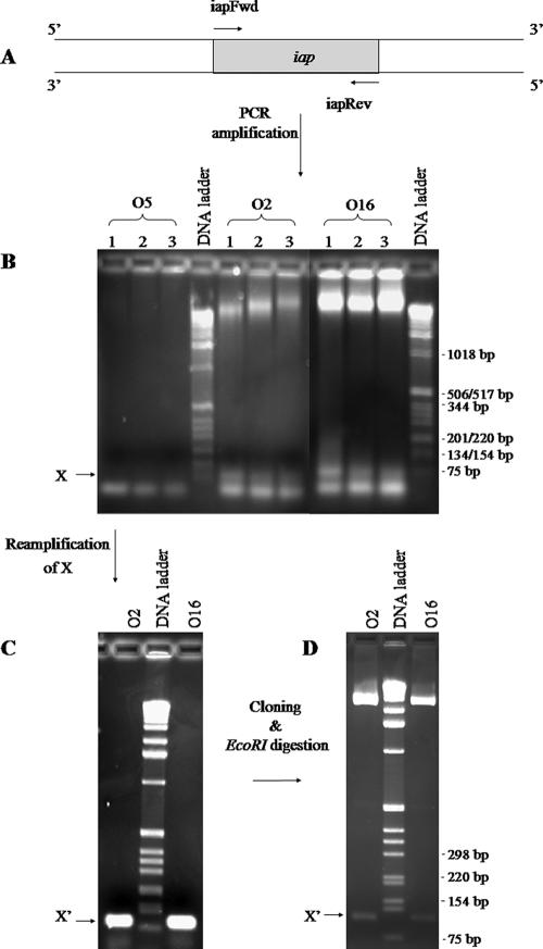
PCR amplification of iap. (A) Primers iapFwd and iapRev were designed to bind to the 5′ and 3′ ends of D3 iap, respectively. (B) Genomic DNAs from serotypes O5, O2, and O16 were used as templates for PCRs. Three different MgSO4 concentrations (1 mM, 1.5 mM, and 2 mM) were used and are indicated by the numbers 1, 2, and 3, respectively. The arrow points to bands corresponding to sizes of ∼95 bp that were present in the O2 and O16 samples but missing in O5. (C) These bands from the O2 and O16 lanes were gel excised and reamplified using primers iapFwd and iapRev. (D) The product, labeled X′, was cloned into a pCR-Blunt II-TOPO vector. Plasmids were extracted from two clones, purified, and digested with EcoRI.

