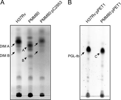
FIG. 3.
TLC analyses of lipids extracted from M. tuberculosis H37Rv and its isogenic Rv2953::Km mutant strain (PMM80). (A) TLC analysis of DIM from the M. tuberculosis wild-type strain, the PMM80 mutant, and the PMM80::pC2953 strain. Lipid extracts dissolved in CHCl3 were separated with petroleum ether-diethylether (90:10, vol/vol), and DIM were visualized by spraying the TLC plate with 10% phosphomolybdic acid in ethanol, followed by heating. Positions of DIM A and DIM B (arrows) and of products A and B (arrowheads) are indicated. (B) TLC analysis of glycolipids extracted from the M. tuberculosis wild-type and PMM80 mutant strains complemented with pPET1. Lipids were dissolved in CHCl3 and run in CHCl3/CH3OH (95:5, vol/vol). Glycoconjugates were visualized by spraying the TLC plate with 0.2% anthrone (wt/vol) in concentrated H2SO4, followed by heating. Positions of M. tuberculosis PGL (PGL-tb; arrow) and product C (arrowhead) are indicated.

