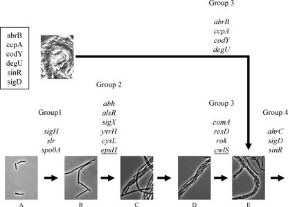
FIG. 6.
Schematic model of cell clustering during pellicle formation. A temporal progression is shown from left to right. Cell cluster formation begins with freely floating planktonic cells (A). Planktonic cells lose motility and form cell chains in a process that appears to be induced by a decrease in the levels of σD-dependent autolysins (B). The number of cell chains increases, and chains come together, forming clusters (C). The clusters of cell chains often make a woven string-like structure (D). Finally, the σH-dependent autolysin CwlS induces cell separation, and the cells in a cluster become clear (E). Genes shown above arrows are required for the indicated stages; epsH and cwlS are underlined. Mutant strains defective in flagellum formation are indicated with a box; these mutant cells form clusters via a different route.

