
Fig. 1.
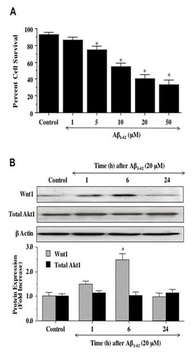
Exposure to Aβ1-42 leads to progressive neuronal injury and is accompanied by an initial increase in expression of Wnt1. (A) Neuronal survival was progressively decreased with increased concentration of Aβ1-42 (*P<0.01 vs. untreated control). (B) Exposure to Aβ1-42resulted in increased Wnt1 expression at 1 hour (h) and significantly at 6 hours (h) following Aβ1-42application, but loss of Wnt1 by 24 hours (h) with expression levels comparable to untreated control (*P<0.01 vs. control). Total Akt1 expression was not altered by Aβ1-42 exposure over a 24 hour course. Each data point represents the mean and ±SEM from 6 experiments.