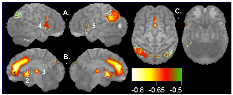
Figure 1.
Decline of brain activity with normal aging: negative correlations between glucose uptake (resting brain activity) and age for a group of 46 healthy subjects (ages 18-90). Surface statistical projection (3D-SSP) of correlation between brain activity and age. Views of the brain: A. Left and right lateral views; B. Medial aspects of left and right hemispheres; C. Dorsal and ventral views. Note large area (1695 voxels) of medial prefrontal cortex including the ACC showing decreased activity with aging. Display threshold is set at r = −0.63 with a significance of p<0.05, corrected for multiple comparisons (586 resels). Local clusters (1, 2, 3; anterior to posterior) shown in medial views. Cluster 2 extends from the subgenual anterior cingulate cortex to the basal forebrain. Cluster 3 in the mediodorsal nucleus of the thalamus is consistent with the metabolic declines in prefrontal cortices. Clusters (4, 5, 6; anterior to posterior) are shown in lateral views. Color scale denotes Pearson correlation coefficient, r, from −0.6 to −0.8. See Table 1 for details.

