
Fig. 7. Spatial selectivity of ACC neurons.
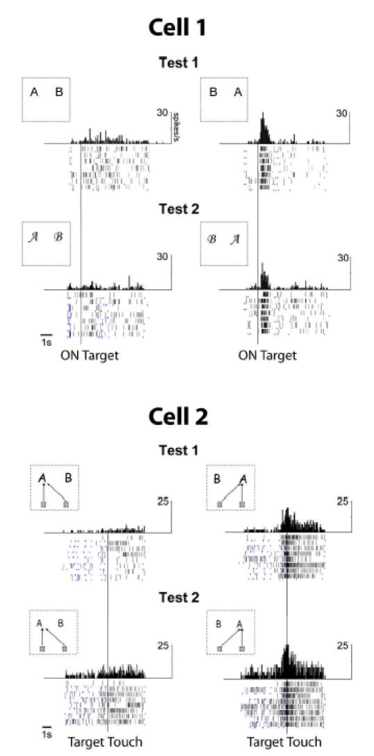
Cell 1 and cell 2 were recorded in the left and right ACC of M1, during the repetition in 2 tests. Positions of letters “A” and “B” in the insets designate the position of the optimal and non-optimal stimulus. Cell 1 is more active when the optimal stimulus is located on the right (Left vs Right, epoch E2: F(1,22)=867.9, p<10−6. Cell 2 is more active when the arm-movements are directed towards the right position (Left vs Right, epoch E4: F(1,31)=239.65, p< 10−6).