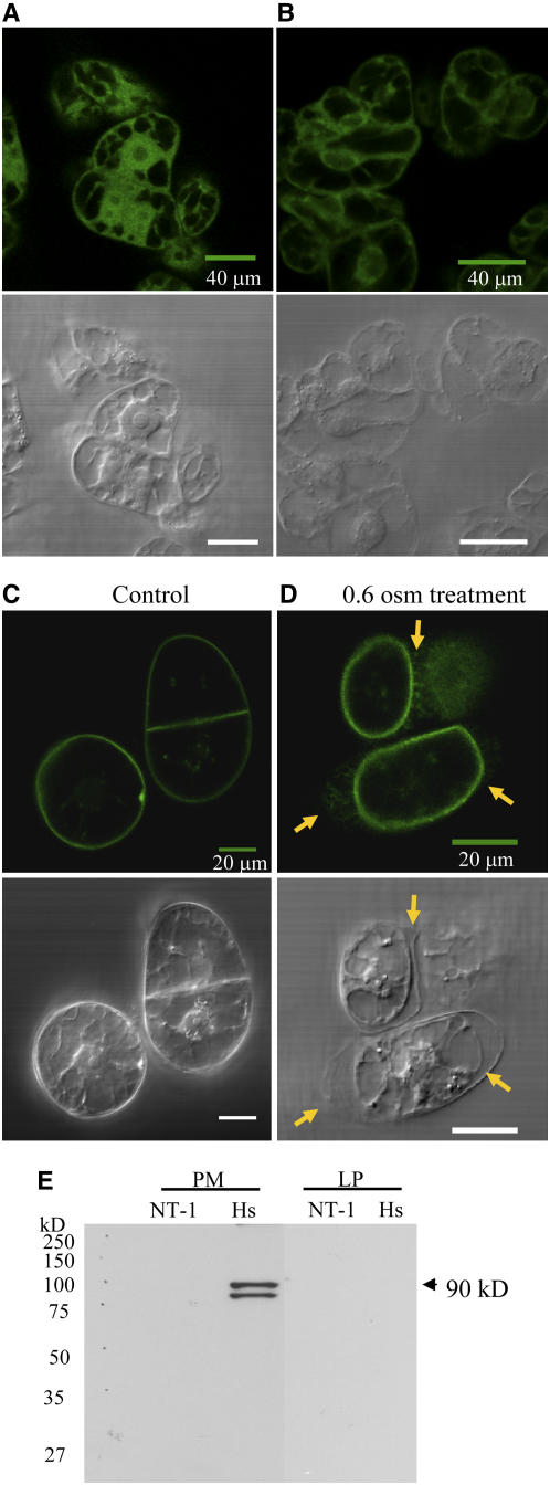
Figure 2.
Full-Length GFP-Hs PIPKIα Localized with the Plasma Membrane.
(A) to (C) Transgenic tobacco cells expressing GFP (A) and GFP-At PIPK10 (B), and GFP-Hs PIPKIα (C) were imaged using a confocal microscope. Top panels show fluorescence, and bottom panels show differential interference contrast images.
(D) To confirm plasma membrane localization of Hs PIPKIα, tobacco cells were treated with 0.6 osmolal sorbitol for 5 min. Arrows indicate the position of the cell wall.
(E) Immunoblot of plasma membrane (PM) and lower-phase (LP) proteins using a monoclonal antibody raised against GFP. The antiserum recognizes a protein of ∼90 kD (the predicted molecular weight of GFP-Hs PIPKIα) and a lower band that may be a proteolytic product in the plasma membrane of Hs PIPKIα cells. Equal amounts of membrane proteins were loaded from NT-1 (wild-type tobacco cells) and Hs (GFP-Hs PIPKIα lines).

