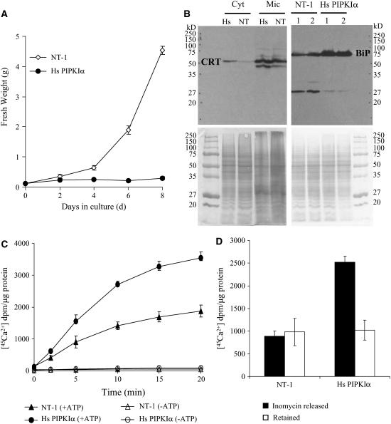
Figure 6.
Hs PIPKIα Lines Did Not Grow in Medium with No Added Calcium and Showed Signs of Changes in Calcium Homeostasis.
(A) Cells (0.25 g/fresh weight) were transferred to 25 mL of fresh medium without added calcium, and fresh weight was monitored every 2 d (wild type, open diamonds; Hs PIPKIα lines, closed circles). The values are averages ± sd of duplicates from two experiments.
(B) Hs PIPKIα cells have increased CRT and BiP. Microsomal membranes (Mic) and cytosolic (Cyt) proteins were prepared from indicated cell lines, and 10 and 30 μg of protein were separated by SDS-PAGE as noted. The calcium binding proteins CRT and BiP were detected with antibodies by immunoblotting as indicated.
(C) Increased Ca2+ uptake in ER from Hs PIPK Iα cells. ER-enriched membrane vesicles were prepared from the wild type (triangles) and Hs PIPK Iα (circles) as previously described (Persson et al., 2001). [45Ca2+] (2 μCi) was added, and uptake was monitored. The ATP-dependent [45Ca2+] uptake (10 μg protein aliquot−1) was measured in the presence (closed symbols) and absence (open symbols) of 3 mM ATP. The radioactivity was measured in a scintillation counter. Data are averages ± sd of duplicates from two independent experiments.
(D) Hs PIPK Iα cells released more ER [45Ca2+]. The Ca2+ ionophore ionomycin (1.5 μM) was added to [45Ca2+]-loaded ER-enriched membrane vesicles (23 min), and membrane vesicles were analyzed for [45Ca2+] after 5 min. Closed bars, [45Ca2+] released; open bars, [45Ca2+] retained by the ER-enriched membranes. The values are averages ± sd of duplicates from two experiments.

