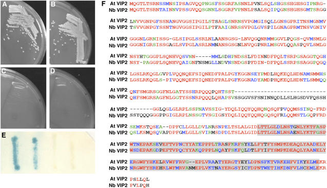
Figure 1.
At VIP2–VirE2 and At VIP2–At VIP1 interactions in the Two-Hybrid System and Amino Acid Sequences of At VIP2 and Nb VIP2.
(A) At VIP2 + VirE2.
(B) At VIP2 + At VIP1.
(C) At VIP2 + human lamin C.
(D) At VIP2 + topoisomerase I.
(E) β-Galactosidase assay. From left to right: At VIP2 + VirE2, At VIP2 + At VIP1, At VIP2 + human lamin C, and At VIP2 + topoisomerase I.
Cells shown in (A) to (D) were grown in the absence of His, Trp, and Leu, and cells shown in (E) were grown in the absence of Trp and Leu.
(F) Multiple sequence alignment by ClustalW (1.81) of amino acid sequences of full-length proteins for At VIP2 and Nb VIP2. The identical amino acids are shown in red, conserved amino acids in blue, semiconserved amino acids in green, and the divergent amino acids in black. The shaded area represents the C-terminal NOT domain between the two proteins.

