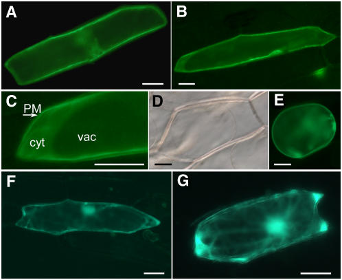
Figure 6.
Transient Expression of GFP-Tagged SCABP8 in Onion Epidermal Cells Visualized by Epifluorescence Microscopy.
(A) Epifluorescence after 20 h of transient expression of SCABP8-GFP.
(B) Epifluorescence after 40 h of transient expression of SCABP8-GFP.
(C) Magnification of the cell shown in (B). PM, plasma membrane; cyt, cytosol; vac, vacuole.
(D) Clear field image of cells plasmolized with 0.5 M sorbitol for 10 min.
(E) Epifluorescence of the same cell shown in (D).
(F) Fluorescence pattern of truncated SCABP8ΔN fused to GFP.
(G) Fluorescence pattern of SCABP8ΔN-GFP.
Bars = 50 μm.

