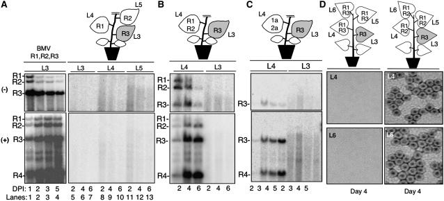
Figure 1.
Reconstitution of BMV RNA Replication in N. benthamiana by Agroinfiltration.
(A) RNA gel blot analysis demonstrating BMV replication for up to 6 d in N. benthamiana. The three lanes labeled “BMV” contain RNAs extracted from leaf tissue where all three BMV RNAs were expressed. The schematics of the plants illustrate the locations where BMV components were infiltrated into each plant. This schematic is intended to denote the location of and the composition of the agroinfiltration and not to denote the relative sizes of the leaves in the plants used. R1, RNA1; R2, RNA2; R3, RNA3. L1 to L6 denote the leaf number. The shaded leaf identifies the location of the RNA3 that will be the focus of the RNA trafficking. Lanes in the image of the RNA gel blots contain RNAs extracted from the leaves shown in the schematic of the plant. The identities of the BMV (+)- and (−)-strand RNAs are indicated to the left of the autoradiogram image. DPI, days after infiltration.
(B) Replication of RNA3 in leaf 4 after agroinfiltration into leaf 3. Leaf 4 was infiltrated with constructs that express RNA1 and RNA2.
(C) RNA3 expressed by agroinfiltration in one leaf replicated in neighboring leaves agroinfiltrated to transiently express the BMV replication proteins.
(D) Production of BMV virions in leaves that reconstituted BMV RNA3 replication. The schematics above the electron micrographs denote the location of BMV components expressed in the leaves relevant to this analysis. The fourth and sixth leaves were used to extract the virions shown in the electron micrographs. The electron micrographs show the presence of the BMV virions (∼28 nm) in leaves 4 and 6.

