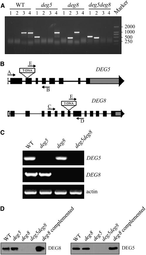
Figure 6.
Identification of the deg5, deg8, and deg5 deg8 Mutants.
(A) PCR analysis of genomic DNA from the wild type and deg5, deg8, and deg5 deg8 mutants to confirm the homozygosity of the mutants. Lane 1, amplification with primers E and B; lane 2, amplification with primers E and D; lane 3, amplification with primers A and B; lane 4, amplification with primers C and D.
(B) Schematic diagram of the DEG5 and DEG8 genes (not to scale). Closed boxes represent exons. Gray boxes represent the untranslated regions of the genes. Arrows indicate the location of primers used for PCR analyses.
(C) RT-PCR analysis of DEG5 and DEG8 gene expression. RT-PCR was performed with the specific primers for DEG5, DEG8, or actin.
(D) Immunodetection of DEG5 and DEG8. Thylakoid membranes were isolated from the wild type, deg5, deg8, and deg5 deg8 mutants, the deg8 mutant complemented with DEG8 cDNA, and the deg5 mutant complemented with DEG5 cDNA and separated by SDS-PAGE. The proteins were immunodetected with anti-DEG5 and anti-DEG8 antibodies.

