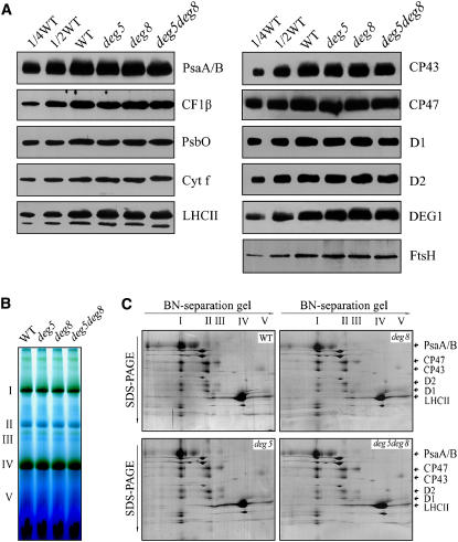
Figure 8.
Analysis of Thylakoid Proteins from deg5, deg8, and deg5 deg8 Mutants and Wild-Type Plants.
(A) Immunoblot analysis of thylakoid proteins from deg5, deg8, and deg5 deg8 mutants and wild-type plants. Thylakoid proteins (1 μg chlorophyll) from the wild type and deg5, deg8, and deg5 deg8 mutants were separated by SDS-urea-PAGE and immunodetected with anti-D1, anti-D2, anti-CP43, anti-CP47, anti-PsbO, anti-LHCII, anti-PsaA/B, anti-cytochrome f, anti-DEG1, anti-CF1β, and anti-FTSH antibodies (which recognize the different isomers of FTSH equally well).
(B) Thylakoid membranes (10 μg chlorophyll) from wild-type and deg5, deg8, and deg5 deg8 mutant leaves were solubilized and separated by BN gel electrophoresis. The positions of protein complexes were identified with appropriate antibodies (see Guo et al., 2005).
(C) BN-PAGE–separated thylakoid proteins were further separated by SDS-urea-PAGE gel. Names of the proteins resolved by the second-dimension SDS-PAGE, previously identified, are indicated by arrows. These experiments were repeated three times independently, and similar results were obtained. Results from a representative experiment are shown.

