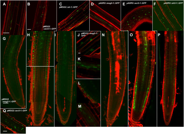
Figure 5.
Expression of mAux/IAA∷GFP transgenes observed with confocal fluorescent microscopy. Longitudinal images of GFP were observed in hypocotyls of 3-d-old etiolated seedlings (A–F) and roots of 1-week-old light-grown seedlings (G–Q) counterstained with 10 μg mL−1 propidium iodide. A, G, J, and K, pMSG2∷msg2-1∷GFP; B, H, and Q, pMSG2∷axr2-1∷GFP; C, I, L, and M, pMSG2∷slr-1∷GFP; D and N, pAXR2∷msg2-1∷GFP; E and O, pAXR2∷axr2-1∷GFP; F and P, pAXR2∷slr-1∷GFP. Images from A to C, E, J, L, and Q are lateral optical sections; the other images are medial sections. J to M and Q, Enlarged images of the central stele in the elongation zone (K and M), and epidermis in the elongation zone (J), the meristematic zone (L), and the root hair initiation zone (Q). Scale bar, 50 μm (A, B, G, and Q) and 10 μm (J). Scales in B to F are the same; scales in G to I and N to P are the same; and scales in J to M are the same.

