Fig. 1.
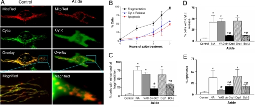
Mitochondrial fragmentation precedes cytochrome c release during apoptosis and is inhibited by dominant-negative Drp1 mutant and Bcl-2. (A) Representative cell images showing mitochondrial fragmentation and cyt.c release after azide treatment. HeLa cells were transfected with MitoRed to fluorescently label mitochondria in red. The transfected cells were subjected to control incubation (Control) or 3 h of 10 mM azide treatment (Azide). Cyt.c was stained in green by indirect immunofluorescence. Images of MitoRed-labeled mitochondria (MitoRed) and cyt.c immunofluorescence (Cyt.c) were collected by confocal microscopy. (B) Time courses of mitochondrial fragmentation, cyt.c release, and apoptosis. HeLa cells transfected with MitoRed were treated with 10 mM azide for indicated time and stained for cyt.c immunofluorescence. Percentages of cells showing mitochondrial fragmentation, cyt.c release, and apoptotic morphology were evaluated by cell counting. Data are means ± SD of three separate experiments. (C–E) Effects of VAD, Drp1, dn-Drp1, and Bcl-2 on mitochondrial fragmentation, cyt.c release, and apoptosis. HeLa cells were cotransfected with MitoRed and dn-Drp1, Drp1, or Bcl-2. The cells were then treated with 10 mM azide for 3 h in the presence or absence of 100 μM VAD. Percentages of mitochondrial fragmentation, cyt.c release, and apoptosis in MitoRed-labeled cells were quantified by cell counting. Data are means ± SD of three separate experiments. *, significantly different from the untreated (Control) group; #, significantly different from the treated no-addition (NA) group.

