Fig. 2.
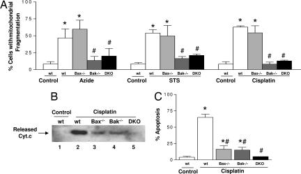
Bak (but not Bax) deficiency blocks mitochondrial fragmentation during apoptosis. Wild-type (wt), Bax-knockout (Bax−/−), Bak-knockout (Bak−/−), and Bax/Bak double-knockout (DKO) MEFs were subjected to apoptotic treatments with 10 mM azide for 3 h, 1 μM STS for 4 h, or 20 μM cisplatin for 16 h. To evaluate mitochondrial fragmentation, the MEFs were transfected with MitoRed before apoptotic treatments. (A) Cells with mitochondrial fragmentation were examined by fluorescence microscopy and quantified by cell counting. (B) To analyze cyt.c release, MEFs after cisplatin treatment were extracted to collect the cytosolic fraction for immunoblot analysis of cyt.c. (C) To analyze apoptosis, MEFs after cisplatin treatment were stained with Hoechst 33342. Apoptosis was evaluated by counting of the cells with typical apoptotic morphology including cellular and nuclear condensation and fragmentation. Data are presented as means ± SD of four separate experiments. *, significantly different from the untreated control group; #, significantly different from the treated wild-type group.

