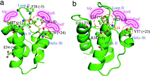
Fig. 2.
Structural models. The convergent mutations were mapped onto structural models of VibBArCP (a) and HMWP2ArCP (b). The proposed differential recognition surfaces are shown in pink. The numbers in parentheses indicates the relative position (RP) of each residue. The position of group I mutations are circled in red.

