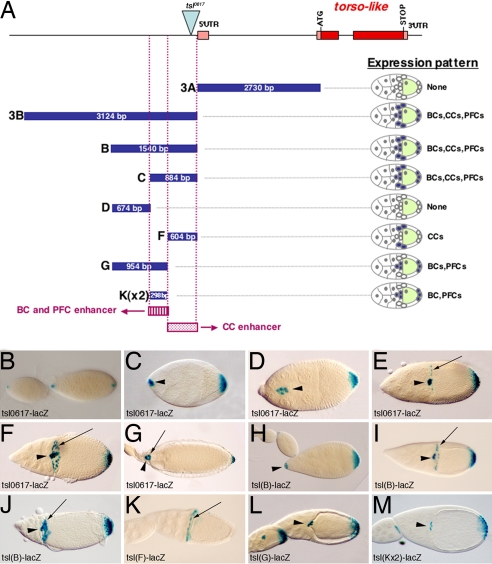
Fig. 1.
Two independent enhancers regulate tsl expression in different cell populations during oogenesis. (A) The tsl genomic fragments tested for enhancer activity by fusion to lacZ reporters. Pink rectangles indicate untranslated exons. Red rectangles indicate translated exons. The blue triangle indicates the P-element in tsl0617. Blue rectangles indicate genomic fragments used for reporter constructs; their size is indicated. Purple rectangles show the identified enhancers. None, No expression. (B–M) X-Gal staining of lacZ reporters. (B–G) tsl0617-lacZ. (B) Expression at stages 7 and 8 of oogenesis in the polar cells at both ends of the egg chamber. (C) At stage 9, expression is first detected in additional PFCs and then in the BCs. (D) At stage 10, expression is detected in the migrating BCs and PFCs. (E) By stage 10B, BCs have reached the oocyte and expression starts to be detected at the CCs. (F) Expression at stage 11. (G) Expression at stage 14. (H–J) tsl(B)-driven expression reproduces all of the tsl pattern [stage 9 (H); stage 10B (I); stage 11 (J)]. (K) tsl(F) drives expression in the CCs. (L) tsl(G) drives expression in the BCs and PFCs. (M) Two copies of the tsl(K) fragment are enough to drive expression in the BCs and PFCs. (K–M) are egg chambers at stages 10B/11. (H–M) These images show ovaries from flies carrying two copies of the construct. Arrows point to CCs, and arrowheads point to BCs. Here and in all images, anterior is to the left.

