
Figure 4.
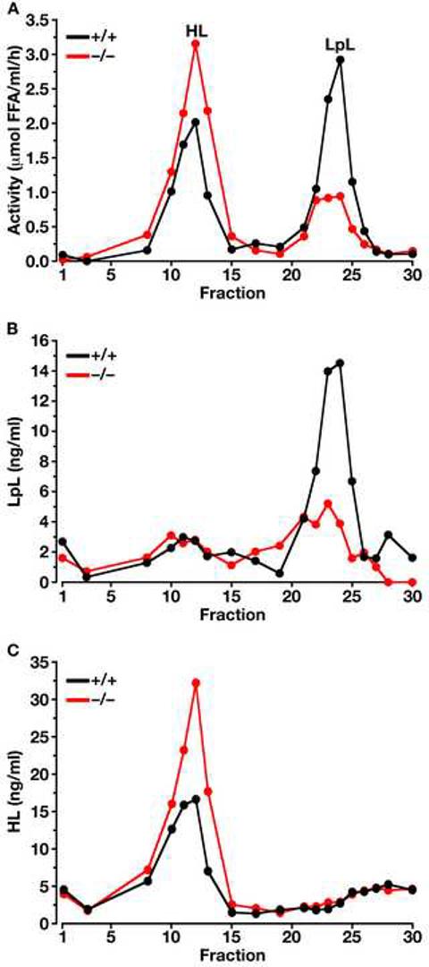
Separation of LpL and HL in the post-heparin plasma of Gpihbp1−/− and wild-type mice on a heparin-Sepharose column. (A) Lipase activities in the different fractions (representative data of four independent experiments). In these studies, the identities of the LpL and HL peaks were verified with immunoassays (B and C, respectively) (Cisar et al., 1989). The specific activity of the LpL in fractions 21–25 of the wild-type and Gpihbp1−/− mouse plasma was not different. Each data point represents the average of duplicate analyses.