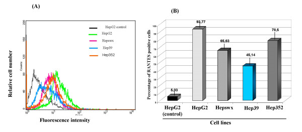
Figure 1.

Immunofluorescence and FACS analysis of RANTES expression. (A) HepG2 cell lines were stained with anti-human RANTES mAb PE conjugated and processed for FACS analysis. (B) Percentage of cells positive for RANTES expression was plotted in columns. HepG2 control indicates parental cell line stained with an antibody isotypically related to anti-RANTES and used as negative control; HepG2 indicates parental cell line, Hepswx vector transfected, Hep39 core expressing cells, Hep352 cell line expressing C-E1-E2-NS2-NS3. One representative experiment out of three independently performed is shown; the standard deviation is indicated by the error bars in B plot; statistical significance (p < 0.05) was determined by Student's t-test for differences.
