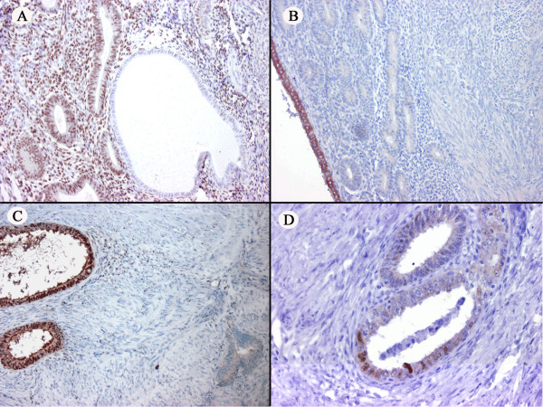
Figure 3.

(A) The atrophic and proliferative component in the endometrium, respectively negative and positive for ER. (B) Aromatase positivity in the superficial layer of a part of the endometrium. (C) The adenomyotic glands throughout the uterus: aromatase expressed by some glands (left), whereas other glands are negative (right) (D) p53 positivity in some adenomyotic glands.
