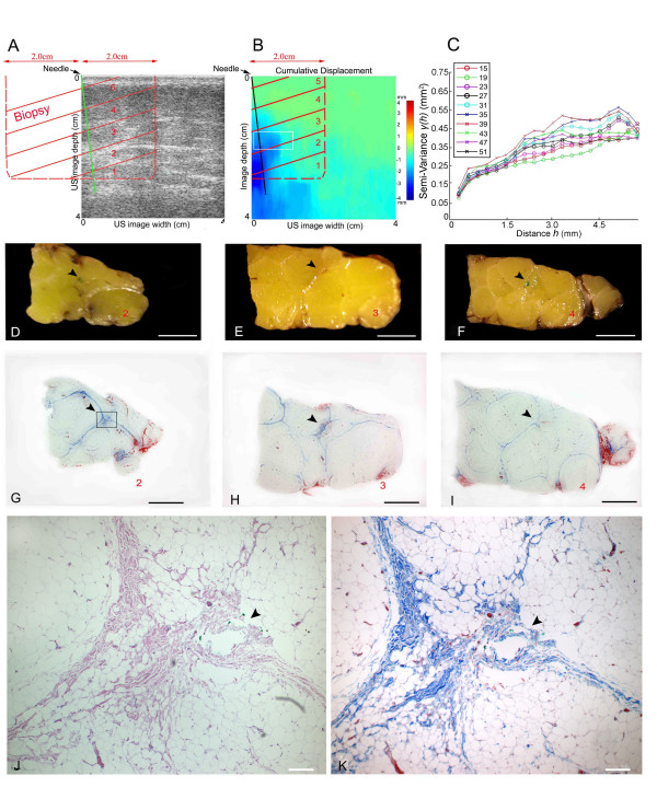
Figure 4.
Effect of needle rotation on connective tissue using ultrasound and histology in a human subject undergoing acupuncture needling on the back during surgery. A: relative locations of ultrasound image, needle and biopsy blocks (labelled 1–5, red letters). B: tissue displacement map during needle rotation shown with ultrasound elasticity imaging showing maximal displacement in the area of the needle. C: semi-variogram analysis of sequential ultrasound frames during needle rotation. Semi-variograms were computed from all ultrasound data points included in the region of interest (10 mm × 5 mm) indicated by white box on panel B. Each semi-variogram line represents one ultrasound frame (frame number shown in inset). D-I: gross (D-F) and microscopic (G-I) images of tissue sections 2,3 and 4 (see location in panels A,B). Arrowheads indicate location of needle (marked with green ink in the tissue specimen). J, K: higher magnification of box area in (G) stained with Hematoxylin/Eosin (J) and Masson Trichrome which stains collagen blue (K). Scale bars: 1 cm (C-I), 0.2 mm (J-K).

