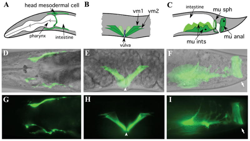
Figure 2. A transcriptional arg-1::gfp reporter is expressed in the head mesodermal cell, the vulval muscles, and the enteric muscles.

The upper panels show line drawings, middle panels show merged micrographs of Nomarski and GFP images, and lower panels show GFP images alone. (A, D, G) the head mesodermal cell, (B, E, H) vm1 and vm2 vulval muscles, and (C, F, I) the four enteric muscles. (F, I) The left intestinal muscle, anal sphincter and anal depressor are visible and the right intestinal muscle is in a different focal plane. The position of the vulval opening is marked by arrowheads and the anal opening is marked by arrows.
