Figure 2.

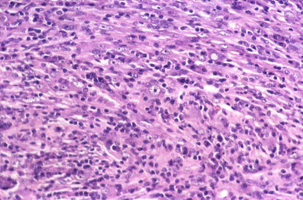
Hematoxylin and eosin staining demonstrating neoplastic proliferation of fusiform cells with pleomorphism and occasional tumour 'giant cells'. These features were typical of inflammatory myofibroblastoma.

Hematoxylin and eosin staining demonstrating neoplastic proliferation of fusiform cells with pleomorphism and occasional tumour 'giant cells'. These features were typical of inflammatory myofibroblastoma.