
Figure 4.
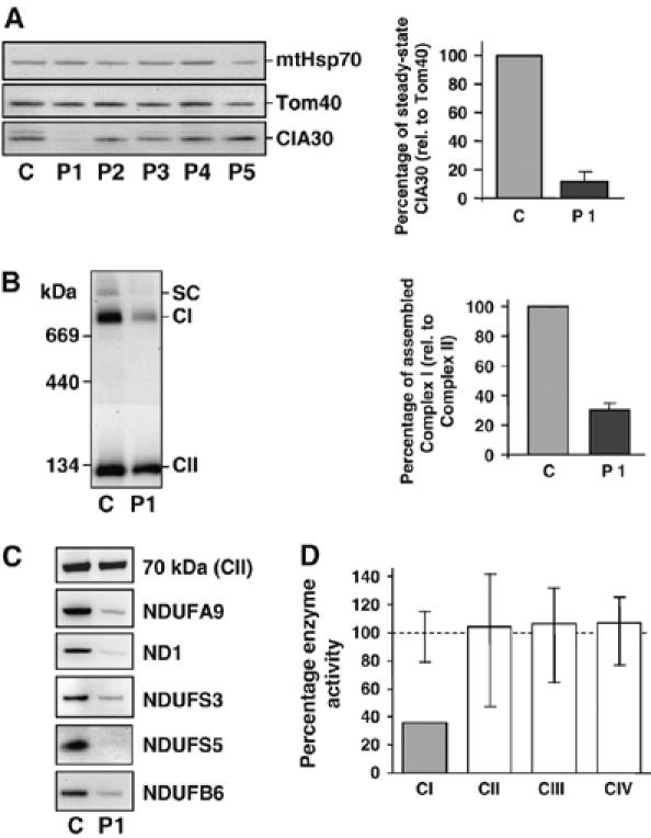
Identification of a complex I-deficient patient cell line with decreased CIA30. (A) Mitochondria isolated from fibroblasts of control (C) and patients (P1–P5) with complex I defects were applied to SDS–PAGE, Western blotted and probed with antibodies against CIA30, Tom40 and mtHsp70 (left panel). Patients had fibroblast complex I activities ranging from 10 to 50% of controls with unknown genetic cause (P2) or with two pathogenic mutations in the complex I subunit genes NDUFV1 (P3), NDUFS4 (P4) or NDUFS6 (P5). The steady-state levels of CIA30 in patient 1 (P1) were quantified against Tom40. Data are mean±s.e.m., n=3 (right panel). (B) Steady-state levels of complex I from patient 1 (P1) fibroblasts were analyzed by BN-PAGE and immunoblotting using antibodies against complex I (CI) and complex II (CII; left panel). SC=complex I/complex III2 supercomplex. The steady-state levels of complex I were quantified against complex II. Data are mean±s.e.m., n=3 (right panel). (C) Mitochondrial extracts from control (C) and patient 1 (P1) fibroblasts were subjected to SDS–PAGE and Western blot analysis against complex I subunits and the complex II 70 kDa subunit (as control). (D) Respiratory chain enzyme activities in patient 1 (P1) lymphoblasts were measured and standardized against the mitochondrial marker enzyme citrate synthase and shown as a percentage of the control mean. Vertical lines represent the observed range for 6 control lymphoblast cell lines.