
Figure 3.
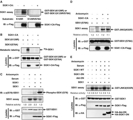
SGK1 phosphorylates SEK1 on serine-78. (A) 293T cells were transfected for 48 h with a vector for Flag-tagged SGK1-CA, and cell lysates were then subjected to immunoprecipitation with anti-Flag antibody. The resulting precipitates were examined for kinase activity with GST-SEK1(K129R) or GST-SEK1(K129R/S78A) as the substrate. Cell lysates were also examined directly by immunoblot analysis with anti-Flag antibody. (B) 293T cells were transfected for 48 h with the indicated combinations of expression vectors for SGK1-CA-Flag, GST-SEK1(K129R), and GST-SEK1(S78A). The transfected cells were metabolically labeled for 3 h with [32P]orthophosphate (100 μCi/ml), after which cell lysates were subjected to immunoprecipitation with anti-GST antibody. The resulting precipitates were analyzed by SDS–PAGE and autoradiography. Cell lysates were also examined directly by immunoblot analysis with antibodies to Flag or to GST. (C) 293T cells were transfected with an expression vector for GST-SEK1, alone or together with a vector for SGK1-Flag. After 48 h of transfection, the cells were deprived of serum for 16 h, and incubated first for 20 min in the absence or presence of 10% FBS and then for 20 min in the absence or presence of 10 μg/ml anisomycin. Cell lysates were subjected to immunoprecipitation with anti-GST antibody, and the resulting precipitates were assayed for SEK1 activity by immune complex kinase assay. Cell lysates were also examined directly by immunoblot analysis with antibodies to phospho-SEK1 (Ser78), GST, or Flag. (D) 293T cells were transfected for 48 h with an expression vector for GST-SEK1(S78A), alone or together with a vector for SGK1-CA-Flag. The cells were then incubated for 20 min in the absence or presence of anisomycin (10 μg/ml). Cell lysates were subjected to immunoprecipitation with anti-GST antibody, and the resulting precipitates were assayed for SEK1 activity by immune complex kinase assay. Cell lysates were also examined directly by immmunoblot analysis with antibodies to GST or to Flag. (E) 293T cells were transfected for 48 h with an expression vector for GST-SEK1, alone or together with vectors for SGK1-Flag and an HA-tagged dominant-negative mutant of either SGK1 (SGK1-KD) or Akt (Akt-DN), as indicated. The cells were deprived of serum for 16 h, and then incubated for 20 min in the absence or presence of 10% FBS. Cells were then left unexposed or exposed to 10 μg/ml anisomycin for 20 min. Cell lysates were assayed for SEK1 activity as for panel C. The cell lysates were also examined directly by immunoblot analysis with antibodies to GST, to Flag, or to HA.