Figure 1.
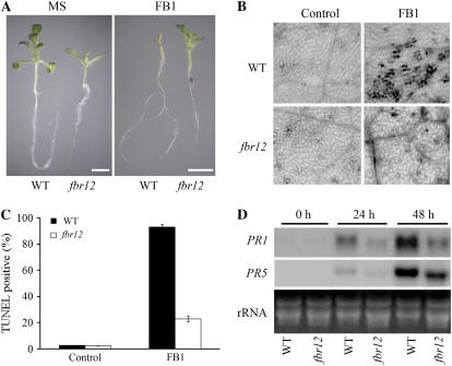
The fbr12 mutant shows an FB1-resistant phenotype and reduced PR gene expression. A, Two-week-old seedlings of wild type (Ws) and fbr12 germinated and grown in Murashige and Skoog medium in the absence or presence of 0.8 μm FB1. Bar = 5 mm. B, Cell death induced by FB1 in wild-type and fbr12 plants. Three-week-old seedlings were treated with dimethyl sulfoxide (DMSO; 0.05%; control) or 2 μm FB1 for 24 h. Leaves were collected and stained with Trypan blue. Black staining represents dead or dying cells. C, Nuclear DNA fragmentation induced by FB1 in wild type and fbr12. Protoplasts prepared from wild-type and fbr12 leaves were treated with DMSO (0.002%; control) or 50 nm FB1 for 12 h and then analyzed by TUNEL staining as described in “Materials and Methods.” TUNEL-positive cells were counted under a fluorescent microscope (n > 1,000 in each experiment). Data presented were mean values of three independent experiments. Bars represent ses. D, FB1-induced expression of PR1 and PR5 genes. Total RNA was prepared from 3-week-old seedlings treated with 3 μm FB1 for various times as indicated and used for northern-blot analysis. Each lane contains 15 μg of RNA. The blot was hybridized with a full-length PR1 or PR5 cDNA probe, respectively.

