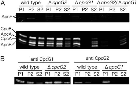
Figure 2.
Localization of PBS proteins in the wild type and cpcG disruptants. Cell extracts were fractionated into low-speed precipitate (P1), high-speed precipitate (P2), and high-speed supernatant (S2). Phycobiliproteins were detected by zinc-induced fluorescence after SDS-PAGE (A), whereas CpcG1 and CpcG2 were detected by immunoblotting (B).

