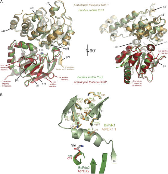
Figure 7.
Modeling the three-dimensional structure of the domains of PLP synthase from Arabidopsis. A, A ribbon representation of PLP synthase from B. subtilis (PDB code 2NV2) is shown in green and is overlaid by a model of PDX1.1 (gold) and PDX2 (red) from Arabidopsis. The two figures show views turned by 90° around a vertical axis in the paper plane. Sequence insertions in the plant enzymes compared to the bacterial enzyme have not been modeled but are indicated in the figure. Visible secondary structure elements have been labeled for reference. B, The part of PLP synthase depicted is proposed to be the route of ammonia transfer. Residues involved in the B. subtilis protein are shown in ball and stick representation (green) overlaid by the equivalent residues of Arabidopsis PDX1.1 (gold). A number of residues are seen to be different in PDX1.1, notably M13 in BsPdx1 is replaced by a Leu.

