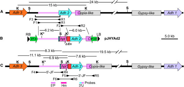
Figure 3.
Strategy for the modification of the Adh locus. A, Genomic structure of the Adh locus containing Adh3, Adh2, Copia- and Gypsy-like retroelements, and Adh1 on chromosome 11. The bracket under the map indicates the 7.0-kb fragment cloned into pINA134 to produce pJHYAd2Ct5 for the authentic 5′-junction fragment in PCR analysis. B, Structure of the vector pJHYAd2. C, Structure of the modified adh2 gene having the hpt and ΔEn sequences inserted in front of its initiation codon. The pink regions represent the homologous sequences that correspond to the flanking Adh2 segments carried by pJHYAd2. Restriction sites S and K indicate SacI and KpnI, respectively, and K* represents the additional KpnI site generated during the pJHYAd2 construction. The horizontal lines and their flanking small arrowheads under the map represent PCR fragments and primers, respectively. The probes 5′P, Hm, and 3′U contain the 0.8-kb Adh2 promoter, the 0.9-kb hpt coding region, and a 0.6-kb segment containing the Adh2-specific 3′ UTR and its 3′ adjacent sequence, respectively. Other symbols are as in Figure 1.

