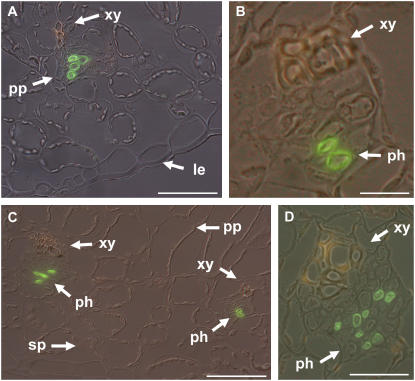
Figure 6.
Immunolocalization of PmPLT1 and PmPLT2 in leaf sections from Plantago plants that had been salt treated for 10 h. A and B, Immunolocalization of PmPLT1 in cross sections of Plantago leaf blades after 10 h of salt stress. A, Overview of a leaf section containing a minor vein. B, Single vein at higher magnification. C and D, Immunolocalization of PmPLT2 in the cross section of a Plantago leaf after 10 h of salt stress. C, Overview of a leaf section containing two minor veins. D, Medium-sized vein at higher magnification. Green labeling shows detection of PmPLT1 (A and B) or PmPLT2 (C and D) with specific antibodies; yellowish staining of xylem vessels results from the autofluorescence of phenolic compounds (le = lower epidermis, ph = phloem, pp = palisade parenchyma, sp = spongy parenchyma, xy = xylem). Scale bars are 50 μm in A, 10 μm in B, 50 μm in C, and 20 μm in D.

